
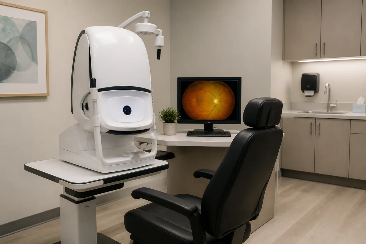
The Optos California is an ultra-widefield retinal imaging system. In a single capture it records a panoramic view of the retina — the light-sensitive tissue lining the back of the eye — reaching far into the periphery that conventional fundus photography cannot show. The resulting image is called an optomap.
How the Imaging Works
You rest your chin on a support and look into the device at a target. Low-powered scanning lasers sweep the retina and build the image in a fraction of a second per eye. Nothing touches the eye, no bright flash is involved, and in most cases dilating drops are not required — which makes the scan practical even when you need to drive yourself home afterwards.
Why We Use It
A great deal of retinal disease begins in the periphery, well outside the area a standard retinal photograph covers. Ultra-widefield imaging helps us find retinal tears and detachments, diabetic retinopathy, vein occlusions, and pigmented lesions earlier than we otherwise might. The California also captures several imaging modes, including colour and autofluorescence, which highlight different layers and reveal changes that colour alone can miss.
What to Expect
The scan is quick, painless, and non-invasive. Because each optomap is stored, we can lay this year's image beside last year's and compare them directly — an objective record of whether something is stable or changing. Your provider will bring the images up on screen and walk you through what they show during the same visit.